
The Biotin‑Avidin System (BAS) is a classic biological bridging and signal amplification platform established in the late 1970s. Leveraging the extremely strong and highly stable specific interaction between biotin and avidin‑family proteins, it breaks the signal ceiling of conventional antigen‑antibody binding. Today, BAS has been widely adopted across immunoassays, in situ labeling, molecular capture, protein purification and many other experimental workflows. Without altering the specificity of antigen‑antibody recognition, BAS functions as an independent "signal relay and amplification module", serving as a well‑established and reproducible technical solution for the detection of trace biomarkers.
Biotin, also known as Vitamin H or Vitamin B7, is a small cyclic carboxylic acid with a molecular weight of only 244 Da. It naturally occurs in animal and plant tissues, serum and cell lysates, acting as a coenzyme for carboxylases to participate in carbohydrate, lipid and amino acid metabolism in vivo.
The biotin molecule consists of an imidazolidone ring and a thiophene ring. The imidazolidone ring serves as the core binding domain for avidin. The terminal carboxyl group on its side chain can be chemically activated into active derivatives such as NHS ester or Sulfo‑NHS ester. Under mild pH conditions, these derivatives covalently couple with amino groups on macromolecules including antibodies, recombinant proteins, peptides and nucleic acids to complete biotin labeling.
Biotin labeling features several key advantages: its small molecular size generally does not significantly impair the antigen‑binding activity of antibodies; the labeling ratio is tunable by adjusting the molar feed ratio to control the number of biotin moieties conjugated to each antibody; labeled products are stable, and free biotin can be readily removed via dialysis, gel filtration or desalting columns to avoid competitive interference with subsequent reactions.
All avidin‑family proteins are homologous tetramers, and each subunit carries an independent biotin‑binding pocket. One protein molecule can bind up to four biotin molecules simultaneously, which constitutes the fundamental structural basis for multivalent bridging and signal amplification in BAS. Nevertheless, the three proteins differ greatly in origin, modification and physicochemical properties, which directly determine background levels and suitable application scenarios.
Extracted from chicken egg white, avidin is a glycoprotein of approximately 64 kDa with ~10% glycosylation and a strongly alkaline isoelectric point (pI≈10). Its glycan chains and positive charges tend to cause non‑specific electrostatic adsorption onto solid phases, tissues and proteins, resulting in high background. It is rarely used directly in modern ELISA.
Derived from Streptomyces, streptavidin has a molecular weight of ~55 kDa without glycosylation and a near‑neutral pI (5~7). Its non‑specific adsorption is far lower than egg avidin, making it the first choice for immunoassays including ELISA, IHC and flow cytometry. Its dissociation constant (Kd) with biotin is around 10‑14 M, representing an affinity far exceeding typical antigen‑antibody interactions. The binding is fast and the complex is exceptionally stable, tolerating wide ranges of pH, temperature, detergents and repeated washing with minimal dissociation.
NeutrAvidin is a deglycosylated variant of egg avidin. It eliminates glycan‑mediated non‑specific binding while retaining the tetrameric biotin‑binding capacity, achieving low background for assays with extremely stringent requirements on non‑specific signals.
The interaction between biotin and streptavidin is non‑covalent yet extraordinarily high‑affinity. Once formed, the complex can barely be disrupted under routine washing and incubation conditions, which underpins the excellent reproducibility and controlled inter‑batch variation of BAS amplification systems.
Based on bridging sequence and reporter placement, BAS has evolved into four mainstream formats with distinct amplification folds, operational complexity and background risks. They serve as the theoretical foundation for kit selection:
Biotinylated antibody + enzyme‑conjugated streptavidin (SA‑HRP / SA‑AP). This workflow is concise with fewer steps, currently the most widely adopted format for BAS‑ELISA. It delivers moderate amplification with manageable background and fits standard sandwich ELISA.
Biotinylated antibody → free avidin → biotinylated enzyme. Avidin acts as a free bridge linking biotin on the antibody and biotin on the enzyme. It offers flexible switching of enzyme reporters, yet requires three incubation steps and is labor‑intensive.
Free avidin and biotinylated enzyme are pre‑mixed at a defined ratio to spontaneously form a mesh‑like, multi‑enzyme enriched ABC complex. One avidin molecule within the complex can recruit multiple biotinylated enzymes, resulting in markedly more concentrated enzyme molecules per binding site and higher amplification gain compared with the BA method. It is suitable for targets of ultra‑low abundance. However, the large complex tends to adsorb non‑specifically onto solid surfaces and raise background. Rigorous optimization of blocking, dilution and incubation time is required. ABC is more common in IHC and cautiously applied in ELISA.
Essentially an improved BA format using glycosylation‑free streptavidin instead of egg avidin, namely the SA‑HRP system widely used in commercial immunoassays, balancing high sensitivity and low background.
- Powerful amplification: the tetravalent binding property enables recruitment of abundant enzyme molecules at a single antigen binding site, converting one binding event into massive enzymatic signals;
- Excellent versatility: a single batch of SA‑HRP reagents is compatible with all biotinylated antibodies. Kit developers avoid generating enzyme‑labeled secondary antibodies individually for each target, simplifying reagent inventory and pairing validation;
- Outstanding reaction stability: biotin‑streptavidin complexes tolerate washing and buffer fluctuations, facilitating tight control of assay CV;
- Modular architecture: BAS acts as an independent amplification unit readily compatible with sandwich, indirect and competitive immunoassays. Existing antigen‑antibody pairs require no redevelopment; sensitivity can be upgraded simply by replacing the detection antibody with its biotinylated version.
- Endogenous biotin interference: serum, plasma and liver/kidney tissue lysates contain native free biotin, which competitively occupies streptavidin binding sites and causes signal suppression (false negative) or abnormal signal elevation (false positive);
- Background risks: ABC complexes, over‑concentrated SA‑HRP and over‑biotinylated antibodies all increase non‑specific adsorption;
- Narrowed linear range: amplified signals are prone to enzymatic saturation, and the high‑concentration end of standard curves readily reaches a plateau;
- Residual free biotin interference: insufficient desalting after antibody biotinylation leaves residual free biotin that consumes SA‑HRP and leads to globally reduced signal intensity.
Conventional ELISA generates signals from directly enzyme‑labeled detection antibodies. Each antibody is conjugated to only a limited number of enzymes. When target proteins are extremely low‑abundance (pg/mL‑level cytokines, chemokines and rare circulating biomarkers), specific signals cannot be distinguished from negative background and the limit of detection fails to meet experimental demands. Embedding BAS as an amplification module into ELISA forms BAS‑ELISA to achieve high‑sensitivity detection.
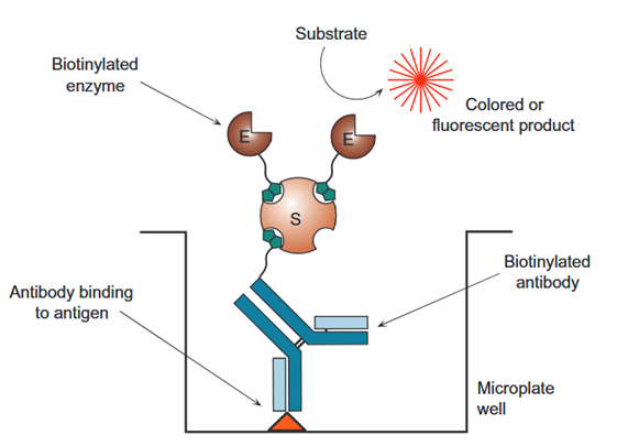
- Coat capture antibody onto the microplate solid phase and block non‑specific binding sites;
- Add test samples; target antigens specifically bind to solid‑phase capture antibodies;
- After washing, add biotinylated detection antibody to form the sandwich immunocomplex: [Solid‑phase capture antibody – antigen – biotinylated detection antibody];
- Wash again and add SA‑HRP (or SA‑AP). Streptavidin binds biotin on the detection antibody and recruits abundant HRP molecules to antigen binding sites;
- Wash away unbound SA‑HRP, add substrates (TMB, pNPP, etc.) for colorimetric or luminescent enzymatic reaction, stop the reaction and read signals via a microplate reader.
Compared with standard sandwich ELISA using directly enzyme‑conjugated secondary antibodies, substantially more enzymes accumulate at each antigen site in BAS‑ELISA. The limit of detection is typically reduced by 5~10 fold, enabling discrimination of weak positive and low‑concentration samples undetectable by regular ELISA.
Beyond sandwich formats, BAS can also be adapted for indirect and competitive ELISA. In indirect ELISA, biotinylated secondary antibodies recognize primary antibodies bound on the solid phase followed by SA‑HRP amplification. In competitive ELISA, biotinylated antigen competes with analyte antigen for solid‑phase antibodies, and binding is traced by SA‑HRP; this format is widely used for small‑molecule toxins and hormone assays.
Low‑abundance cytokines (TGF‑β, IL family, IFN, VEGF, etc.), trace tumor markers, neural injury biomarkers, autoantibodies, neutralizing antibody titers, drug residues and exosome‑associated protein quantification for research and in vitro diagnostics. This system is compatible with colorimetric and chemiluminescent plate ELISA, and can also be transferred to western blot, multiplex immunofluorescence, bead‑based flow capture and other immunoassay platforms.
- Prioritize streptavidin conjugates over egg avidin to minimize glycan‑related background;
- Free biotin must be removed by desalting after antibody biotinylation;
- Evaluate interference for serum samples rich in endogenous biotin; implement blocking steps if necessary;
- Test gradients of SA‑HRP working concentration and incubation time to avoid elevated background from excessive enzyme loading;
- Set low‑concentration gradients for standard curves to assess LOD and LOQ, and anticipate signal saturation at high analyte levels.