
Within the phosphatidylinositol signal cascade, cell-surface receptors bind external signaling ligands to trigger phospholipase C (PLC) activation. The activated PLC enzyme then breaks down phosphatidylinositol-(4,5)-bisphosphate (PIP2), splitting it into two distinct secondary messenger molecules: inositol trisphosphate (IP3) and diacylglycerol (DAG). These two messengers jointly translate extracellular stimulation into intracellular signaling events, forming the phosphatidylinositol signal transmission network, which is also widely referred to as the dual messenger system.
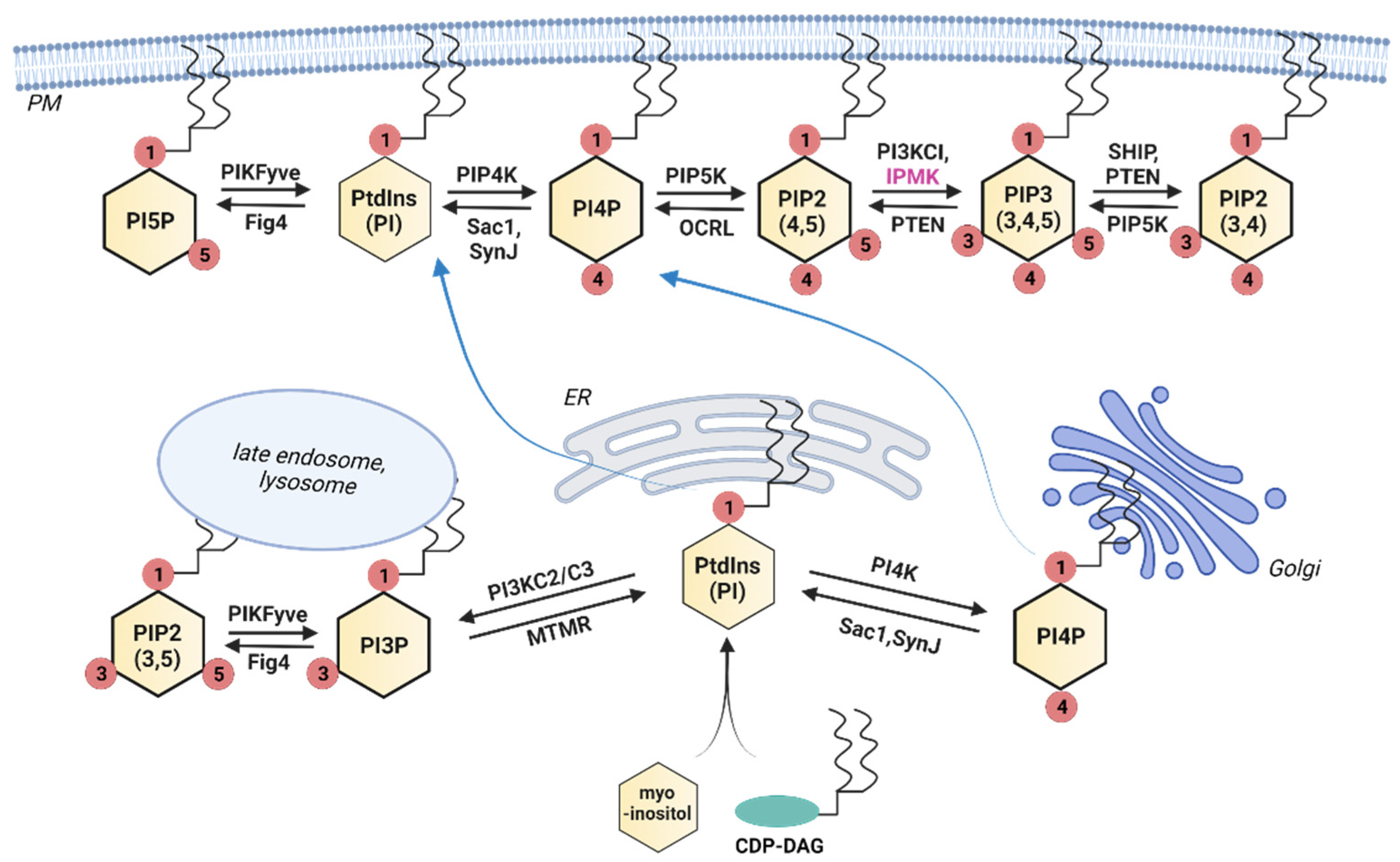
Lipid kinase-mediated phosphorylation of phosphatidylinositol generates a range of phosphoinositide derivatives, including PI3P, PI(4,5)P2 and PI(3,4,5)P3. These phosphoinositide lipids act as vital signaling intermediates that engage multiple downstream cascades, among which the PI3K-Akt axis governs core cellular processes including proliferation, viability maintenance and metabolic regulation. Beyond this major pathway, phosphoinositide-dependent signaling also coordinates diverse cellular behaviors, such as cell migratory movement, endocytic vesicle formation and plasma membrane remodeling.
The core functional components of this signaling network include membrane receptors, heterotrimeric G proteins and downstream effector enzymes. Among all effector subtypes identified so far, PLC-β has been the most comprehensively characterized and deeply researched. Extracellular bioactive substances such as hormones and neurotransmitters can associate with pathway-specific cell-surface receptors, thereby initiating a sequential chain of intracellular signal transduction.
When hormonal or other extracellular signaling agents attach to G protein-coupled membrane receptors, conformational shifts occur in coupled G protein complexes, which in turn stimulates plasma membrane-localized PLC-β. Catalytically active PLC subsequently mediates the cleavage of membrane-resident PIP2 to produce IP3 and DAG.
IP3 molecules interact with calcium ion-gated channels located on the endoplasmic reticulum (ER) membrane, opening these pores to drive efflux of stored Ca²⁺ from the ER lumen. After binding free Ca²⁺ ions, calmodulin (CaM) undergoes structural rearrangement and becomes capable of activating calmodulin-dependent protein kinases. Triggered CaM-kinase activity modulates numerous downstream target proteins and initiates calcium-dependent signal cascades, which control a broad spectrum of cellular physiological events: inflammatory reactions, metabolic turnover, programmed cell death, cytoskeletal movement, gamete fertilization, short- and long-term memory formation, and immune defensive responses.
Separately, calcium ions discharged from the ER raise cytoplasmic calcium concentrations, prompting protein kinase C (PKC) to translocate from the cytosol to the inner leaflet of the plasma membrane. At this membrane location, DAG functions as a co-factor to fully activate PKC, hence Ca²⁺ is sometimes described as the tertiary messenger in this cascade. Once activated, PKC catalyzes phosphorylation on numerous substrate proteins, triggering divergent cellular outcomes including secretory vesicle release, smooth muscle contraction, and the proliferation or differentiation of cell populations.