
A research paper titled HSF1 promotes CD69+Treg differentiation to inhibit colitis progression was recently published in the journal Theranostics. This research verified that CD69 knockdown triggers the onset of autoimmune illnesses including inflammatory bowel disease. Moreover, heat shock factor 1 (HSF1) elevates CD69 transcription levels to drive the differentiation of CD69-positive regulatory T cells (CD69+Tregs), a process indispensable for Tregs to exert immune-suppressive functions. For patients suffering from autoimmune diseases, elevating the quantity of CD69+Treg cells is expected to effectively alleviate clinical symptoms.
As the earliest surface molecule upregulated upon lymphocyte activation, CD69 serves as a core immune regulatory factor with essential physiological functions. Accumulating research data confirms that CD69 exerts crucial regulatory effects in hematological disorders and autoimmune conditions. Nevertheless, its complete biological functions and detailed immune regulatory mechanisms remain to be further explored and validated. This article systematically elaborates on this key molecular target from multiple dimensions.
Also named AIM, EA-1, Leu23 and MLR-3, CD69 is a type II transmembrane glycoprotein selectively expressed on activated lymphocytes. Human-derived CD69 is composed of 199 amino acid residues, consisting of three distinct structural segments: a 40-amino-acid intracellular domain, a 21-amino-acid transmembrane region, and a 138-amino-acid extracellular domain. The extracellular segment contains a C-type lectin domain featuring a calcium-dependent carbohydrate recognition domain (CRD), classifying CD69 into the C-type lectin receptor superfamily. Serine and threonine residues at the intracellular terminus can undergo phosphorylation modification, and this molecule is capable of binding to CD23 and CD72 simultaneously.
CD69 is also a constituent component of the NK cell-specific genetic factor complex. Unlike NKR-P1, Ly-49, NKG2 and other molecules exclusively restricted to NK cells, CD69 exhibits widespread expression across multiple cell populations, endowing it with versatile biological functionalities.
CD69 is a cell-surface antigen that only gets transcribed and translated after lymphocyte activation. Subsequent research demonstrates that nearly all blood cell types can be induced to express CD69 under appropriate stimulation. The inducible cell spectrum covers T lymphocytes, B lymphocytes, NK cells, monocytes, neutrophils, eosinophils and thymocytes, among others. Owing to its exclusive expression in activated cells rather than resting ones, CD69 has become an ideal phenotypic marker to identify activated immune cells.
During T cell activation, CD69 acts as a typical activation marker alongside CD25 (IL-2 receptor) and CD71 (transferrin receptor). Beyond biomarker applications, CD69 also delivers co-stimulatory signals to participate in immune activation cascades. Experimental evidence indicates that CD69-positive T cells display markedly elevated mRNA levels of IL-12β, IL-18α and IL-16β; when supplemented with recombinant IL-12 or IL-18, these cells further secrete large quantities of interferon-γ (IFN-γ). Recent advanced studies additionally reveal that CD69 participates in cell apoptosis regulation in parallel with its activation-promoting effects.
Although the definitive specific ligand of CD69 has not been fully identified, existing studies confirm its binding interaction with Galectin-1 (Gal-1), through which it modulates the differentiation and function of Th17 cells. Overall, CD69 cross-linking initiates intracellular signal cascades and triggers diversified immune responses, exerting extensive regulatory impacts on the biological behaviors of hematopoietic cells.
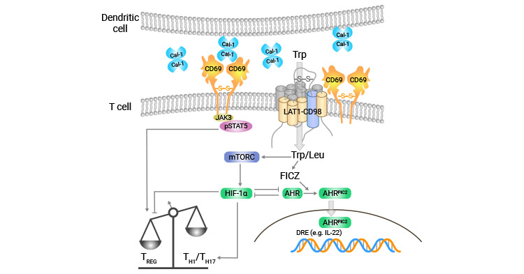
Quiescent T cells do not express detectable CD69. Stimulation via anti-CD3/TCR complex, CD2 or CD28 ligation, or treatment with PKC agonists including phorbol myristate acetate (PMA) and phytohemagglutinin (PHA) will robustly induce CD69 upregulation on T cell surfaces.
The intact CD3/TCR complex is a prerequisite for CD69-initiated T cell activation. In addition, PMA activates GTP-binding proteins and Ras to engage downstream intracellular signaling axes, which is essential for triggering CD69 expression in T lymphocytes.
Resting NK cells are also CD69-negative. Cross-linking CD16, or stimulation with IL-2, IL-7, IL-12, IFN-α or PMA, activates NK cells and induces CD69 expression. This process relies on multiple kinases including diverse PKC isoforms and protein tyrosine kinases (PTKs) to coordinate multiple signaling pathways. The cytotoxic capacity of CD69-activated NK cells is negatively modulated by CD94. Previous reports further confirm that CD69 activates extracellular signal-regulated kinase (ERK), whereas the inhibitory receptor CD94/NKG2-A counteracts this ERK activation process.
For splenic and lymph node-resident B cells, lipopolysaccharide (LPS) serves as an effective inducer for CD69 expression. In in vitro culture systems, peripheral blood eosinophils (EOS) upregulate CD69 upon exposure to IL-2, IL-3, IL-5, IL-13, GM-CSF, IFN-γ and other inflammatory cytokines.
In vivo experiments verify tight connections between CD69 and programmed cell death. T cells highly expressing CD69 after in vivo activation are prone to rapid spontaneous apoptosis when cultured ex vivo. Notably, CD69-mediated apoptosis differs substantially from CD95-triggered apoptosis: CD95 drives apoptosis across nearly all cell types, while CD69-induced cell death is largely limited to activated T cells, eosinophils and monocytes.
Four independent signaling axes contribute to CD69-initiated monocyte apoptosis: (1) nitric oxide (NO)-dependent pathway; (2) phospholipase A2/oxidized phospholipid cascade pathway; (3) pertussis toxin (PTX)-sensitive G protein-dependent pathway; (4) CD69-induced TGF-β synthesis further accelerates apoptotic progression.
Caspase family proteins are the central executors of apoptosis. In the human eosinophil cell line EOL-1, incubation with monoclonal antibodies targeting CD45, CD45R, CD45RB, CD95 or CD69 significantly boosts apoptosis; this phenotype can be rescued by Caspase-8 and Caspase-9 inhibitors. CD69 cross-linking activates Caspase-3 and Caspase-8 sequentially. Anti-CD69 monoclonal antibodies induce eosinophil apoptosis and downregulate anti-apoptotic Bcl-2 expression, yet exogenous GM-CSF stabilizes intracellular Bcl-2 levels. Even in GM-CSF-cultured CD69-positive cells, CD69 still reduces Bcl-2 abundance and triggers apoptosis, solidifying the close linkage between CD69-mediated apoptosis and Bcl-2 protein regulation.
Apoptosis triggered by mature T cell activation predominantly relies on the FAS signaling pathway. Tonsillar activated T cells with high CD69 expression display drastically elevated spontaneous apoptosis rates in vivo. Cytokines such as CXCL12/SDF-1, TIL16, IL-16, IL-17, IL-12 and IL-15 can suppress this apoptotic tendency, whereas phosphatidylinositol 3-kinase (PI3K) inhibition accelerates cell death. These findings illustrate that multiple exogenous signals jointly tune the apoptotic susceptibility of in vivo activated CD69+ T cells.
In lymphoma, acute/chronic leukemia, multiple myeloma and other hematological malignancies, peripheral blood CD4+ and CD8+ T lymphocytes exhibit significantly higher surface levels of activation markers CD69 and CD71, indicative of persistent T cell hyperactivation in these disease states.
Acute myeloid leukemia (AML) is characterized by aberrant proliferation and blocked differentiation of hematopoietic stem cells, with massive accumulation of immature leukemic blasts that occupy normal hematopoietic niches, ultimately leading to anemia, recurrent infection and hemorrhage. Downregulated CD69 expression represents one critical pathogenic factor for AML progression.
Reduced CD69 levels curtail the secretion of IL-2, TNF-α and other key cytokines, hinder effector T cell generation and impair systemic anti-tumor immune responses. Clinically, CD69 expression rebounds sharply in AML patients who achieve complete remission after chemotherapy, confirming sustained T cell activation suppression and compromised immune function in untreated AML patients.
Chronic lymphocytic leukemia (CLL) arises from malignant clonal expansion of CD5+CD19+ B lymphocytes. Upon mitogen stimulation in vitro, B cells isolated from CLL patients show comparable CD69 expression to healthy controls, accompanied by defective CD95 expression patterns. Further validation reveals that CLL patient cells overexpress surface activation antigens including CD23, CD25, CD69 and CD71; specifically, CD69 expression intensity positively correlates with peripheral blood lymphocyte count and unfavorable clinical staging, serving as an independent prognostic biomarker.
Monocytes isolated from myelodysplastic syndrome (MDS) patients feature prominent CD69 upregulation, which correlates with systemic immune dysfunction. Excessively high monocyte CD69 expression impairs pathogen clearance capacity, rendering MDS patients more vulnerable to infectious complications.
Current CD69-related research focuses heavily on systemic lupus erythematosus (SLE), rheumatoid arthritis (RA) and other autoimmune illnesses. Clinical cohort data shows elevated peripheral blood CD69 levels in autoimmune disease patients, which decline markedly following effective intervention; CD69 expression intensity positively correlates with disease activity and progression severity.
SLE is defined by disrupted functional homeostasis of T and B lymphocytes. Under identical stimulation conditions, lymphocytes from SLE patients show stronger CD69 upregulation and moderate proliferative responses compared with healthy donors. No significant CD69 expression differences exist between CD4+ and CD8+ T cell subsets in SLE patients, proving CD69 mediates early activation signal transmission in both T cell populations.
Synovial fluid CD4+ and CD8+ T cells from RA patients both show robust CD69 overexpression, confirming full activation of these two T cell subsets. When comparing synovial fluid samples from RA, seronegative spondyloarthropathy and crystal-induced arthritis patients, the percentage of CD69-positive lymphocytes is comparable across groups, yet RA patients display the highest CD69 expression per positive cell. Additionally, lymphocyte CD69 expression level in RA synovial fluid is positively correlated with local IL-15 concentration.
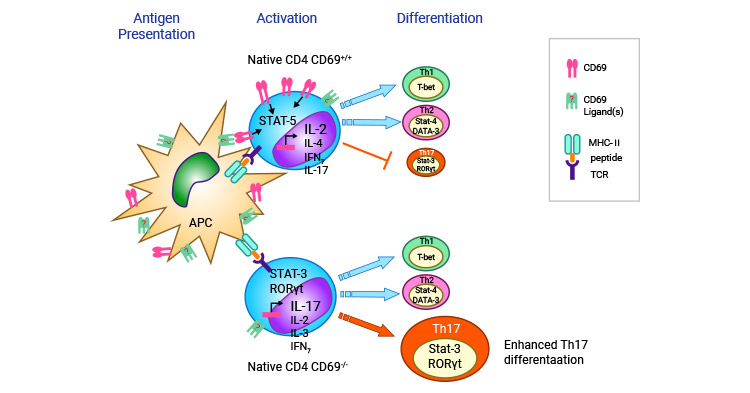
In a murine autoimmune myocarditis model, CD69 negatively regulates myocardial inflammatory injury by modulating cardiac-specific Th17 cell populations, which are major drivers of myocarditis and numerous inflammatory pathologies. In vivo mechanistic assays confirm CD69 restrains T cell differentiation toward Th17 lineage via the Jak3/Stat5 signaling axis.
Coronary heart disease (CHD) patients present elevated serum levels of CD69, DKK-1, IL-10, IL-6, total cholesterol (TC), triglyceride (TG) and low-density lipoprotein (LDL), with marker levels rising in parallel with worsening coronary artery stenosis. Asthma patient airway-resident T cells express CD69 upon activation; activated CD69 further induces T cell apoptosis by modulating TGF-β synthesis and secretion, offering novel therapeutic ideas for asthma management.
For solid tumors including gastric carcinoma, hepatocellular carcinoma and colorectal cancer, peripheral blood CD69 expression is significantly decreased and negatively associated with tumor staging. Surgical resection or chemotherapy for primary lesions restores CD69 expression to near-normal levels in most patients.
GeneFrontier Corp. is developing the anti-CD69 monoclonal antibody GFC-101, with potential therapeutic applications for irritable bowel syndrome, rheumatoid arthritis and respiratory inflammatory diseases. As an early activation-triggering molecule, CD69 initiates cascade immune responses, making CD69-targeted antibodies and small-molecule modulators promising drug candidates under active development. In CLL treatment, blocking B cell receptor signaling to suppress CD69 expression may improve therapeutic responses in patients insensitive to bendamustine monotherapy. Multiple novel potential CD69 ligands, including Galectin-1 and S1PR1, have been identified in recent years. With continuous deepening of basic mechanistic research on CD69, more innovative CD69-targeted immunotherapies will be translated into clinical practice for hematological and autoimmune disease treatment.