
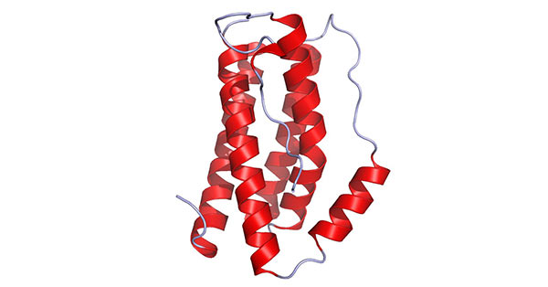
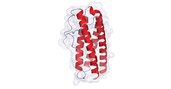
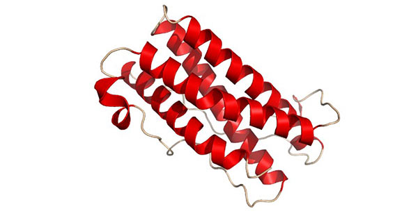
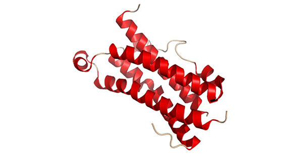
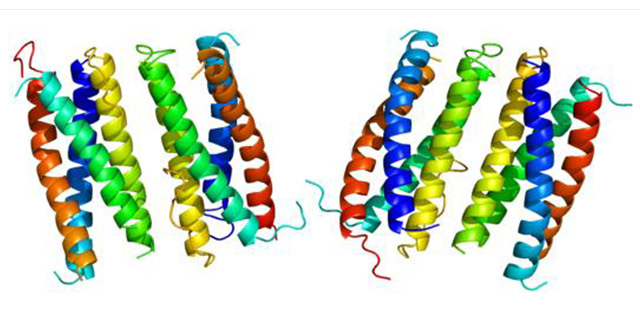
IL-6 is classified as polypeptide substance, composed of two glycoprotein subunits. The alpha subunit has a molecular weight of 80 kDa, while the molecular weight of beta subunit reaches 130 kDa. The alpha subunit lacks intracellular functional domain, only combining with IL-6 at low affinity. After forming preliminary complex, it will rapidly bind to high-affinity beta subunit, and further transmit biological signals into cells via beta subunit. The spatial conformation of IL-6 presents four long alpha helix chains, arranged in an up-up-down-down topological structure.
Abnormal sustained secretion of IL-6 will trigger the occurrence and deterioration of autoimmune disorders and chronic inflammatory illnesses. Initially named B cell stimulating factor-2, this molecule can promote activated B cells to generate antibodies. When cooperating with TGF-β, IL-6 facilitates naive CD4+ T cells to differentiate into Th17 cells, and meanwhile restrains the formation of regulatory T cells induced by TGF-β.
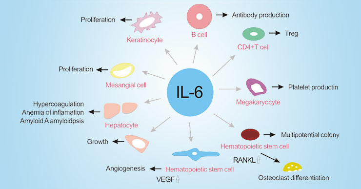
IL-6 can induce hepatocytes to synthesize acute phase reactive proteins such as C-reactive protein, fibrinogen, serum amyloid A and hepcidin, and suppress albumin synthesis simultaneously. Excessive serum amyloid A and hepcidin will separately lead to amyloidosis and inflammatory anemia. Within bone marrow tissue, IL-6 accelerates megakaryocyte maturation into platelets and activates hematopoietic stem cells. Besides, it promotes osteoclast differentiation, angiogenesis, proliferation of keratinocytes and mesangial cells, and accelerates growth of myeloma and plasmacytoma cells.
This cytokine exerts multiple physiological effects. It boosts hematopoietic development and stimulates hepatocytes to secrete acute phase reactive proteins. Moreover, it inhibits adipocyte formation, activates osteoclast activity, changes neuronal phenotypic characteristics, accelerates tissue fibrosis, and regulates biological behaviors of chondrocytes, synoviocytes and B lymphocytes.
IL-27 activates downstream JAK/STAT signaling cascade by combining with receptor complex formed by IL-27α and gp130. High expression of IL-27 can be detected in lesion sites of rheumatoid arthritis, psoriatic arthritis, systemic sclerosis, inflammatory bowel disease, multiple sclerosis and sarcoidosis, proving its close linkage with inflammatory pathological changes.
IL-31 conducts biological signals through compound receptor made up of IL-31RA and OSMRβ, which are expressed on immune cells and epithelial cells. The activated downstream pathways include ERK1/2 MAP kinase pathway, PI3K/AKT pathway and JAK1/2 pathway.
Gene sequence of LIF shows high conservation between human and mouse species. Mature LIF exists as monomeric glycoprotein with frequent glycosylation modification. Unglycosylated protein weighs 20–25 kDa, while glycosylated form ranges from 37–63 kDa in molecular weight.
The full-length protein contains 196 amino acid residues. Sequence comparison confirms OSM has close evolutionary relation with other gp130 family cytokines, sharing 22% sequence identity and 30% similarity with LIF. OSM and LIF genes are tandemly arranged on human chromosome 22, owning highly consistent gene structure, promoter sequence and intron-exon arrangement.
CNTF is one of the most deeply researched members among cytokines acting via gp130/LIFRβ receptor compound. Genetic mutation causing LIFRβ functional loss will lead to severe clinical symptoms, which indicates these family cytokines undertake irreplaceable physiological tasks during individual growth and development.
Primary structural analysis proves CNTF belongs to intracellular soluble protein, featuring classic four antiparallel helix bundle conformation. It binds with specific CNTF receptor first, then recruits gp130 and LIF-R to assemble complete signal complex and activate subsequent biological responses.
CT-1 can be detected in normal adult lung tissue, as well as fetal and adult airway smooth muscle cells. It activates gp130 dependent signal transmission and stimulates JAK/STAT pathway, delivering hypertrophy induction and cell protection signals inside cardiomyocytes. Meanwhile, CT-1 also presents obvious neurotrophic activity.
Similar to CNTF and CT-1, NP can sustain embryonic motor neuron survival, promote neural precursor proliferation and accelerate astrocyte differentiation in vitro. Its expression peak appears when other IL-6 family cytokines stay at low level, reflecting unique physiological roles in mouse nervous system development.
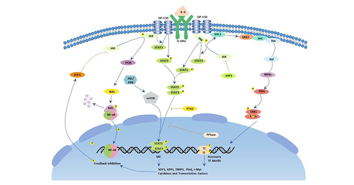
Signal activation starts from the combination between IL-6 and its specific receptor IL-6R. IL-6R has two existing forms on cell surface: membrane anchored type and soluble free type. Cells without membrane IL-6R can still receive stimulation from compound formed by IL-6 and soluble IL-6R. After binding specific receptor, IL-6 further recruits two gp130 molecules to assemble tetramer or hexamer compound structure. The activated gp130 subsequently initiates two core downstream signaling cascades: JAK/STAT pathway and Ras/MAPK pathway.
In JAK/STAT pathway, activated JAK kinase catalyzes phosphorylation modification of STAT3 and SHP2 transcription factors. Phosphorylated STAT3 forms dimer complex and transfers into cell nucleus, binding with target gene promoter segments to start gene transcription. STAT3 is essential for gp130 mediated cell survival and cell cycle transformation. c-Myc and Pim serve as downstream target genes, jointly maintaining cell survival and cycle progression. SHP2 connects cytokine receptor and Ras/MAPK cascade, acting as key mediator of cell proliferation signal.
For Ras/MAPK pathway, activated gp130 facilitates assembly of Shc-Grb2-Sos complex and activates Ras protein. Active Ras sequentially triggers phosphorylation level elevation of downstream MAPK related kinases, regulating cell growth state. Major phosphorylated substrate proteins include c-Myc, c-Jun and c-Fos.
Mass clinical research verifies IL-6 can be used as effective diagnostic biomarker for various illnesses. Relevant studies demonstrate IL-6 affects differentiation tendency of CD4 T cells, participating in the occurrence and advancement of autoimmune diseases. Elevated IL-6 concentration will aggravate inflammatory lesions, and partial patients show enhanced cellular response toward this cytokine.
Apart from inflammatory disorders, abnormal IL-6 expression is also closely associated with malignant tumors, covering gastrointestinal carcinoma, pancreatic cancer and colorectal cancer.
[2] Yoshida H, Nakaya M, et al. Interleukin 27: a double-edged sword for offense and defense. J Leukoc Biol, 2009.
[3] Fearon U. Interleukin-27: A master regulator in inflammation. Arthritis & Rheumatism, 2011.
[4] Pflanz S, Timans JC, et al. IL-27, a heterodimeric cytokine. Immunity, 2002.
[5] Shahrara S, Huang Q, et al. TH-17 cells in rheumatoid arthritis. Arthritis Res Ther, 2008.
[6] Jones BE, Maerz MD, et al. IL-6: a cytokine at the crossroads of autoimmunity. Curr Opin Immunol, 2018.