
Human cytomegalovirus (CMV) has parasitized human hosts across the entire course of human evolution and currently poses infection risks to most populations globally. This pathogen is closely linked to a wide spectrum of human disorders, including various malignant tumors, cardiovascular lesions, gastrointestinal ailments, and unexplained ophthalmic pathological conditions.
Human cytomegalovirus (also termed HCMV or human herpesvirus type 5, HHV-5) is a double-stranded DNA virus falling under the Betaherpesvirinae subfamily of the Herpesviridae family. It represents a pervasive infectious agent that invades 50% to 80% of individuals at some stage of their lifespan, yet seldom triggers apparent clinical symptoms. Pathological conditions correlated with HHV-5 infection encompass infectious mononucleosis and interstitial pneumonia. Following initial invasion, the virus can enter a dormant latent state and regain activity under specific internal and external stimuli. HCMV solely transmits between human carriers, with no transmission routes involving food, drinking water, or animal vectors.
HCMV features a distinctive triple-layer structural architecture, consisting of an outermost lipid bilayer envelope, an intermediate amorphous tegument layer, and an inner icosahedral capsid that wraps the viral genetic material. Four core components constitute the HCMV capsid framework: major capsid protein (MCP/pUL86), triplex dimer (TRI2/pUL85), triplex monomer (TRI1/pUL46), and minimal capsid protein (SCP/pUL48A).
The HCMV genome is split into two unique coding segments, designated Unique Long (UL) and Unique Short (US). These two segments are flanked by terminal and internal repeat sequences, arranged in the fixed order TRL-UL-IRL-IRS-US-TRS.
Two key glycoprotein complexes embedded on the viral envelope mediate host cell recognition and entry:
- Trimer complex (gH/gL/gO): Interacts with corresponding surface receptors to facilitate infection of fibroblast cells
- Pentamer complex (gH/gL/UL128-131): Targets specific receptors to invade epithelial and endothelial cells
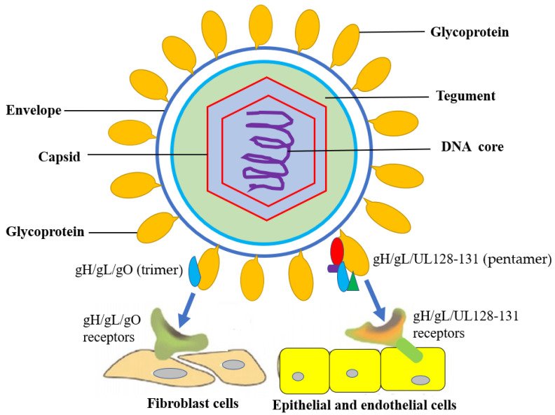
Figure 1. Structural schematic diagram of HCMV
The complete life cycle of HCMV consists of multiple sequential biological stages: viral adhesion and host cell penetration, viral gene expression and genome duplication, viral particle assembly, genome encapsidation, virion maturation, and eventual release out of infected host cells.
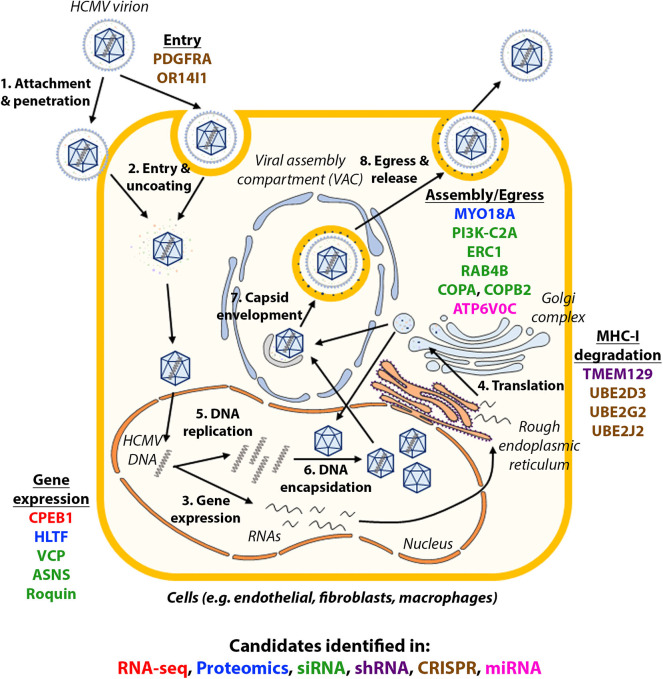
Figure 2. Schematic diagram illustrating the whole HCMV life cycle
HCMV infects fibroblast cells through membrane fusion reactions mediated by the trimeric gH/gL/gO (UL75/UL115/UL74) complex, which binds platelet-derived growth factor receptor α (PDGFRα) on cell surfaces. TGFβRIII and neuregulin-2 (NRG2) function as auxiliary receptors for this trimer complex, boosting the entry efficiency within fibroblasts. The surface gM/gN (UL100/UL73) dimer and gB (UL55) trimer are both indispensable for viral entry. The gM/gN dimer facilitates viral attachment via interaction with heparin sulfate proteoglycans on cell membranes, while the gB trimer acts as a core mediator to trigger membrane fusion.
For epithelial and endothelial cell infection, HCMV adopts a distinct invasion pathway relying on low-pH dependent endocytosis. This process depends on the binding between pentamer envelope glycoprotein complex gH/gL/UL128/UL130/UL131 and cell surface neuropilin-2 (Nrp2). Multiple other surface molecules including CD147, CD46, and OR14I1 have also been verified to participate in pentamer-mediated epithelial cell entry.
After crossing the host cell membrane barrier, tegument-coated viral capsids are transported to nuclear pores along microtubule filaments, driven by the binding between tegument proteins and host microtubule transport machinery. The viral genome is subsequently released into the host nucleus through nuclear pore channels. Certain tegument proteins such as pp65 (pUL83) and pp71 (pUL82) detach from capsids and enter the nucleus independently. pp65 participates in regulating viral replication progress, whereas pp71 mainly controls the activation of viral gene expression.
The incoming linear viral genome serves as dual templates: one for de novo transcription to generate viral protein precursors, and another for the synthesis of progeny viral genomes. Inside the host nucleus, linear HCMV DNA undergoes cyclization and aggregates into concatemeric DNA chains. The tegument protein pp71 binds Daxx, a host death domain-associated protein that recruits histone deacetylases to suppress transcription. This binding triggers proteolytic degradation of Daxx, thereby removing transcriptional inhibition and activating the expression of immediate early (IE) genes. Protein products translated from IE genes interact with cellular cytokines to further activate the transcription of early (E) and late (L) genes in sequence. Early viral proteins are primarily responsible for viral transcriptional regulation and genome replication, while late proteins constitute nearly all structural components of mature virions.
HCMV encodes six core protein factors essential for viral genome replication: UL54 (DNA polymerase catalytic subunit), UL44 (DNA polymerase processivity auxiliary factor), UL70 (primase), UL105 (helicase), UL102 (primase-associated cofactor), and UL57 (single-stranded DNA-binding protein).
The transcription of late viral genes initiates capsid assembly processes within the host cell nucleus. Once capsid structural assembly finishes, a multi-subunit terminase complex composed of pUL51/pUL52/pUL56/pUL77/pUL89/pUL93 attaches to concatemeric viral DNA, cutting long-chain genomes into unit-length fragments for packaging into immature procapsids to form complete nucleocapsids.
After viral DNA replication and genome packaging into nascent capsids, DNA-containing nucleocapsids depart from nuclear replication compartments and cross the nuclear envelope through a cyclic envelopment-deenvelopment-reenvelopment transport mechanism. This process includes primary envelopment at the inner nuclear membrane (INM) and de-envelopment upon passing the outer nuclear membrane (ONM). Afterwards, incomplete nucleocapsids acquire additional tegument proteins and final lipid envelopes after transportation to cytoplasmic virion assembly compartments (vAC) derived from Golgi organelles. Fully mature infectious virions are then secreted out of host cells via membrane budding.
HCMV is capable of infecting diverse human cell lineages, covering epithelial cells, endothelial cells, fibroblasts and myeloid precursor cells. Two distinct infection phenotypes can be induced by HCMV: lytic productive infection and latent persistent infection, whose conversion is governed by the activity status of viral major immediate early promoter (MIEP). During lytic infection, the MIEP region is decorated with transcriptional activation epigenetic markers, which drives high-level expression of core lytic IE genes. The lytic replication cycle follows an ordered cascade of IE, E and L gene expression, eventually generating large quantities of progeny infectious virions.
Following initial lytic infection, HCMV can establish permanent latent infection inside host cells, enabling the virus to evade clearance by the host immune system for decades. To date, latent HCMV has only been identified in early myeloid lineage cells, including CD34+ hematopoietic stem cells (HSCs), monocyte progenitor cells and peripheral blood monocytes. Within latently infected cells, no progeny virions are produced; the MIEP locus is modified with repressive chromatin markers to silence the whole viral genome. Viral reactivation mainly originates from latent CD34+ hematopoietic progenitor cells (HPCs) and CD14+ monocytes, which frequently occurs after allogeneic blood transfusion and solid organ transplantation.
For healthy immunocompetent individuals, primary HCMV infection usually induces mild or even zero clinical symptoms. In contrast, congenital infected infants and immunosuppressed patients such as organ transplant recipients will suffer severe pathogenic damage triggered by HCMV.
Primary HCMV infection spreads mainly through intrauterine vertical transmission, breastfeeding, or direct contact with virus-laden secretions including saliva and genital secretions. The detailed transmission approaches between infected and uninfected individuals via mucosal contact are summarized below:
- Direct contact with infected saliva or urine, especially secretions from infants and young children
- Transmission through intimate sexual contact and genital fluid exchange
- Newborn infection via breast milk during lactation
- Vertical transmission from pregnant women to fetuses during gestation, or neonatal infection during childbirth
- Iatrogenic infection via transplanted organs and allogeneic blood transfusions
Organ transplant recipients face remarkably higher infection risks, since long-term immunosuppressive drugs are administered to prevent immune-mediated graft rejection.
During primary HCMV exposure, host immune cells synthesize abundant specific antibodies targeting multiple viral antigens, covering tegument structural proteins, envelope glycoproteins and non-structural functional proteins. Existing research data prove that humoral antibody immunity plays a critical role in limiting viral amplification and alleviating clinical pathological manifestations.
After invading target host cells, abundant HCMV gene products are expressed to interfere with natural killer (NK) cell-mediated recognition, helping the virus escape immune surveillance and subsequent elimination. At least 12 kinds of HCMV-encoded proteins are confirmed to modulate NK cell function, including US20, UL16, UL17, UL18, UL40, UL43, UL140, UL83, UL141-UL144, and UL148. Protein products encoded by UL16, UL17, UL40, UL140, and UL142 simulate the structure of host HLA class I molecules to inhibit NK cell cytotoxic activity. Besides protein regulators, HCMV microRNA miR-UL122 can also suppress the surface expression of host MICB to block NK cell recognition signals.