
Blog

A Systematic Introduction to Coagulation: Factors, Cascade and Clinical Implications
Coagulation, or blood clotting, describes the process through which liquid blood transforms into a gel-like state to form a clot. This process is often referred to as secondary hemostasis, representing the second phase in stopping bleeding from damaged blood vessels. The first phase, primary hemostasis, involves vasoconstriction and platelet aggregation at the injury site. The coagulation mechanism relies on platelet activation, adhesion and aggregation, as well as fibrin deposition and maturation. Coagulation disorders can lead to excessive bleeding or pathological clot formation known as thrombosis.
1
The Function of Coagulation
Coagulation is a biological process that converts blood into a solidified state. Its key functions mainly include two aspects: sealing injuries to the skin, which serves as the body’s primary barrier against external pathogens, and preserving the integrity of the circulatory system to ensure oxygen and nutrient delivery to tissues. This process is also present in many non-human animals and can be regarded as a natural protective mechanism similar to a biological bandage.
2
Coagulation Factors
Blood clotting is a sequential cascade involving a group of plasma components known as coagulation factors. In total, there are 12 major coagulation factors numbered with Roman numerals from I to XIII. You may notice that factor VI is absent from the list; this is because factor VI, also called factor Va, is now recognized as the activated form of factor V and is no longer assigned an independent number.
Coagulation factor I, also known as fibrinogen, is a glycoprotein synthesized in hepatocytes. It consists of two trimers, each formed by three distinct polypeptide chains: fibrinogen α chain (encoded by the FGA gene), fibrinogen β chain (encoded by the FGB gene), and fibrinogen γ chain (encoded by the FGG gene). In addition, coagulation factor XIII is a 320 kDa glycoprotein tetramer composed of two A subunits and two B subunits.

Table 1. The family of coagulation factors

NumberName / AliasChemical EssenceSynthetic siteInvolving pathwayFunction
IFibrinogenglycoproteinLiverFinal common pathwayForms clot (fibrin)
IIProthrombinglycoproteinLiverFinal common pathwayIts active form (IIa) activates I, V, X, VII, VIII, XI, XIII, protein C, platelets
IIITissue factorglycoproteinTissue
Endotheliocyte
monocyte
Tissue factor pathwayCo-factor of VIIa (formerly known as factor III)
IVCalciumcalcium ion-Three pathwaysRequired for coagulation factors to bind to phospholipid (formerly known as factor IV)
Vproaccelerin, labile factorglycoproteinLiverFinal common pathwayCo-factor of X with which it forms the prothrombinase complex
VIIstable factor, proconvertinglycoproteinLiverTissue factor pathwayActivates IX, X
VIIIAntihemophilic factor AglycoproteinLiverContact activation pathwayCo-factor of IX with which it forms the tenase complex
IXAntihemophilic factor BglycoproteinLiverContact activation pathwayActivates X; forms tenase complex with factor VIII
XStuart-Prower factorglycoproteinLiverFinal common pathwayActivates II; forms prothrombinase complex with factor V
XIplasma thromboplastin antecedentglycoproteinLiverContact activation pathwayActivates IX
XIIHageman factorglycoproteinLiverContact activation pathwayActivates factor XI, VII and prekallikrein
XIIIfibrin-stabilizing factorglycoproteinLiver, plateletFinal common pathwayCrosslinks fibrin
3
Coagulation Cascade
As the term suggests, a cascade refers to a multi-step physiological process in which each step triggers the next, leading to signal amplification once initiated. The coagulation cascade begins with either the contact activation pathway or the tissue factor pathway, then converges into the final common pathway to produce a fibrin clot.
The contact activation pathway responds to abnormalities within blood vessel walls in the absence of direct tissue damage. In contrast, the tissue factor pathway is initiated by actual tissue injury. Although these two pathways start differently, they eventually merge into the common pathway. Both rely on a series of coagulation factors that circulate in an inactive state until activated during the cascade. 
Tissue Factor (Extrinsic) Pathway
Also known as the extrinsic pathway, the tissue factor pathway is the more common and well-characterized arm of the coagulation cascade. When tissue injury occurs and bleeding begins, circulating factor VII binds to tissue factor (TF) expressed on damaged cells, forming an active TF-VIIa complex. This complex then activates factors IX and X. Factor Xa produced in this reaction marks the entry point into the combined common pathway.
Contact Activation (Intrinsic) Pathway
The intrinsic pathway is triggered when blood interacts with collagen in damaged vessel walls, leading to the assembly of a complex involving high-molecular-weight kininogen (HMWK), prekallikrein, and factor XII. This promotes the conversion of factor XII into its active form, XIIa. Factor XIIa then activates factor XI to XIa, which in turn activates factor IX to IXa. Hemophilia B is an inherited bleeding disorder caused by a deficiency in factor IX. Activated factor IXa converts factor X to Xa, at which point the intrinsic pathway joins the common pathway.
Final Common Pathway
In the final common pathway, factor Xa catalyzes the conversion of prothrombin (factor II) into thrombin (IIa). Thrombin activates platelets and promotes their accumulation at the injury site to form a platelet plug. Von Willebrand factor (vWF) supports platelet adhesion to damaged vessel walls. A deficiency in vWF causes von Willebrand disease, the most common inherited bleeding disorder.
4
The Related Disease of Coagulation
Coagulation abnormalities may result in bleeding, thrombosis, or in some cases both, depending on the specific defect. This section describes several representative disorders linked to coagulation dysfunction, including coagulation factor disorders, platelet disorders, inflammatory conditions, and other related diseases.
Coagulation Disorders
Coagulation disorders refer to impairments in the body’s ability to regulate blood clot formation. The most well-known example is hemophilia, in which the lack of a critical coagulation component leads to prolonged bleeding. Many other coagulation disorders arise from diverse causes. Studies have shown that cancer patients frequently exhibit coagulation abnormalities due to tumor progression, impaired myelopoiesis, hypoproteinemia, endothelial dysfunction, and organ dysfunction caused by metastasis. Clinical correlations between coagulation disorders and tumor growth have also been increasingly reported.
Current standard care for hemophilia involves prophylactic intravenous infusions two to three times per week, which imposes a considerable burden on patients. Research by Swystun LL and colleagues has also explored gene therapy strategies for coagulation disorders.
Platelet Disorders
Platelet disorders include three main categories: elevated platelet count (thrombocythemia and reactive thrombocytosis), reduced platelet count (thrombocytopenia), and impaired platelet function. Platelets are small cytoplasmic fragments derived from bone marrow that play a central role in hemostasis and wound healing.
Low platelet counts increase the risk of bleeding ranging from mild to severe, while extremely high counts may raise the risk of thrombosis. Platelet dysfunction refers to qualitative defects in platelet activity. For instance, in von Willebrand disease, platelets fail to properly aggregate or adhere to vessel walls, leading to abnormal bleeding. Recent studies indicate that high-throughput sequencing enables rapid diagnosis of inherited platelet disorders.
Inflammation
More than 150 years ago, pathological studies noted excessive coagulation activation during inflammatory responses. However, the molecular mechanisms linking these two systems have only been clarified in recent decades. Although often studied separately, coagulation and inflammation are closely integrated and tightly regulated systems with extensive crosstalk that shapes the body’s response to injury and infection. Several key molecular interactions show potential as therapeutic targets for inflammatory and thrombotic diseases.
Tissue Factor
During injury or infection, circulating monocytes and subendothelial cells such as smooth muscle cells are activated through ROS, cytokines, endotoxins, and DAMPs acting on pattern recognition receptors including TLRs. Tissue factor becomes exposed to circulating blood and its expression is sustained by inflammatory mediators released by leukocytes, platelets, endothelial cells, and perivascular cells.

TF initiates the activation of factor VIIa, factor Xa, and thrombin (IIa), which in turn trigger platelet activation and PAR-mediated signaling. This leads to increased cytokine release, upregulated adhesion molecules such as ICAM-1 and VCAM-1, and reduced expression of vasculoprotective molecules such as thrombomodulin. The release of P-selectin, vWF, PF4, and CD40L further promotes platelet and leukocyte recruitment and the formation of neutrophil extracellular traps (NETs). Thrombin also contributes to complement activation, generating C5a and C5b-9, which amplify inflammation and coagulation.
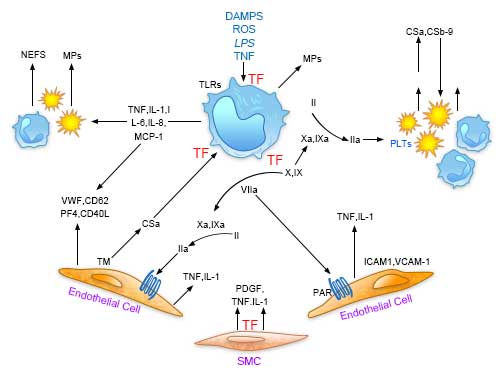
Central role of tissue factor (TF) in the interplay between coagulation and inflammation

Fig. 1. Central role of tissue factor (TF) in the interplay between coagulation and inflammation.


Factor XII of coagulation
Upon contact with negatively charged surfaces, factor XII undergoes autoactivation to form FXIIa. FXIIa then interacts with high-molecular-weight kininogen (HK) bound to prekallikrein (PPK). FXIIa converts PPK into kallikrein, which further accelerates FXII activation and promotes the release of bradykinin from HK. Bradykinin binds to endothelial and neutrophil receptors to trigger inflammatory responses.

FXIIa also activates factor XI in the coagulation cascade, promoting additional thrombin generation. Free thrombin drives both coagulation and inflammation, while thrombin bound to thrombomodulin promotes the production of activated protein C (APC) and TAFIa. C1-inhibitor serves as a major negative regulator of FXIIa, FXIa, and kallikrein.
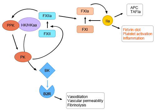
Factor XII activation at the nexus of coagulation and inflammation

Fig.2. Factor XII activation at the nexus of coagulation and inflammation.
5
Clinical significance
Coagulation is a highly dynamic process, and advances in understanding the coagulation system have influenced modern anesthetic practice. This section introduces two clinically relevant applications related to coagulation.
Coagulation tests
Coagulation testing is a routine laboratory examination with minimal risks and side effects. A blood sample is collected and analyzed in a clinical laboratory. Conditions associated with coagulation abnormalities include liver disease, thrombophilia, and hemophilia. Coagulation tests are widely used to monitor patients taking anticoagulant medications and are often recommended preoperatively.
Common coagulation assays include complete blood count (CBC), factor V assay, fibrinogen level measurement, prothrombin time (PT / PT-INR), platelet count, thrombin time, and bleeding time.
Thrombin-antithrombin complex
The coagulation system is a precisely regulated network that generates thrombin in a controlled manner. It includes zymogens, enzymes, cofactors, and inhibitors that ensure rapid thrombin production at injury sites while limiting excessive activity in healthy circulation.
Thrombin-antithrombin complex (TAT) is a small molecular complex that contributes to the inactivation of multiple coagulation enzymes. Antithrombin, a component of TAT, is a 432-amino-acid glycoprotein produced in the liver.
The half-life of the thrombin-antithrombin complex in plasma is approximately three days. Under normal physiological conditions, the concentration of thrombin-antithrombin complex in human plasma is around 0.12 mg/mL. Protein purification and cDNA sequencing conducted on thrombin-antithrombin complex isolated from various species have revealed that key amino acid residues are located at the sixth position of the peptide chain. Three intramolecular disulfide bonds are formed among six monomeric amino acids, specifically Cys8‑Cys128, Cys21‑Cys95, and Cys248‑Cys430.
Four potential N‑glycosylation sites exist within the primary structure of thrombin-antithrombin complex, corresponding to asparagine (Asn) residues at positions 96, 135, 155, and 192; similar glycosylation sites are also highly conserved in antithrombin across other species. In the predominant isoform of thrombin-antithrombin complex, all four glycosylation sites are covalently modified with oligosaccharide side chains, giving this isoform a molecular weight of up to 58,200 Da. In the secondary isoform of thrombin-antithrombin complex, however, the potential N‑glycosylation site at asparagine 135 remains unoccupied.
   💬 WhatsApp