
DNA can be described as an intricate, tightly coiled biological cable carrying life’s genetic information. Its remarkable packaging capability relies entirely on histones, which compact the 2-meter-long DNA strand into a tiny 6-micron eukaryotic cell and securely store all genetic data.
Albrecht Kossel first identified histones back in 1884. Histones represent a category of alkaline proteins residing inside eukaryotic cell nuclei and act as the primary structural building blocks of chromosomes. These proteins contain abundant lysine and arginine residues, granting them positive electrical charges. This characteristic enables them to tightly bind negatively charged DNA strands to form chromatin complexes.
Histones fall into two major groups: core histones and linker histones. Core histone subtypes include H2A, H2B, H3 and H4, while linker histones consist of H1 alongside its homologous protein H5. Within eukaryotic cells, core histones are generally synthesized concurrently with DNA replication, with their expression levels peaking during the S phase of the cell cycle.
Core histones exhibit extremely high evolutionary conservation across all species, ranging from yeast to human beings. Each core histone possesses two key structural regions: a C-terminal histone-fold domain (HFD) responsible for assembling histone octamers, and an unstructured N-terminal tail that extends outward from the nucleosome core particle. This protruding tail undergoes diverse post-translational modifications (PTMs).
The nucleosome core particle is built around an octamer unit: one (H3-H4)₂ tetramer sandwiched between two separate H2A-H2B dimers. Roughly 147 base pairs of DNA wrap twice around this octamer complex. When linker histone H1 binds to the nucleosome alongside the extra DNA segments at the DNA entry and exit points of the core particle, the classic "beads on a string" chromatin structure forms, laying the foundation for higher-order chromatin architectures. Six nucleosome units combined with H1 linker histones assemble into a solenoid fiber. These solenoid fibers further coil around protein scaffolds, and repeated rounds of coiling ultimately form the complete chromosomal matrix.

Figure 1. Histone structure and nucleosome assembly
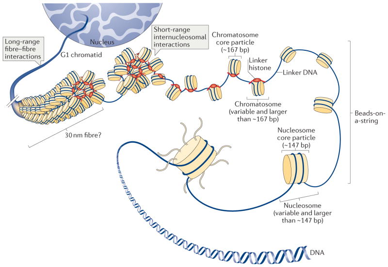
Figure 2. Multiple hierarchical layers of chromatin folding and histone architecture
Histone variants refer to slightly altered isoforms of histone proteins, most commonly variants of core histones. Though they share core structural features with canonical histones, they carry unique biochemical traits and biological roles. These variants alter nucleosome physical properties—such as the length of DNA wrapped around the complex or the overall stability of nucleosomes—to generate structural diversity. Collectively, histone variants expand chromatin structural complexity and enable sophisticated epigenetic regulation of the eukaryotic genome.
The table below catalogs all human histone proteins and their corresponding variants:
| Super family | Family | Subfamily | Members |
|---|---|---|---|
| Linker Histones | H1 | H1F | H1F0, H1FNT, H1FOO , H1FX |
| H1H1 | HIST1H1A , HIST1H1B , HIST1H1C , HIST1H1D , HIST1H1E , HIST1H1T | ||
| Core Histones | H2A | H2AF | H2AFB1 , H2AFB2/3, H2AFJ , H2AFV , H2AFX, H2AFY, H2AFY2, H2AFZ |
| H2A1 | HIST1H2AA , HIST1H2AB/E , HIST1H2AC , HIST1H2AD , HIST1H2AG, HIST1H2AI, HIST1H2AJ, HIST1H2AK, HIST1H2AL, HIST1H2AM | ||
| H2A2 | HIST2H2AA3, HIST2H2AC | ||
| H2B | H2BF | H2BFM , H2BC12L, H2BFWT | |
| H2B1 | HIST1H2BA , HIST1H2BB , HIST1H2BC/E/F/G/I, HIST1H2BD , HIST1H2BH , HIST1H2BJ, HIST1H2BK, HIST1H2BL, HIST1H2BM, HIST1H2BN , HIST1H2BO | ||
| H2B2 | HIST2H2BE | ||
| H3 | H3A1 | HIST1H3A , HIST1H3B, HIST1H3C, HIST1H3D, HIST1H3E, HIST1H3F, HIST1H3G, HIST1H3H, HIST1H3I, HIST1H3J | |
| H3A2 | HIST2H3C | ||
| H3A3 | HIST3H3 | ||
| H4 | H41 | HIST1H4A , HIST1H4B, HIST1H4C, HIST1H4D, HIST1H4E, HIST1H4F, HIST1H4G, HIST1H4H, HIST1H4I, HIST1H4J, HIST1H4K, HIST1H4L | |
| H44 | HIST4H4 |
Data within this table is extracted from Wikipedia
For decades, histones were merely viewed as inert packaging materials for nuclear DNA, until researchers uncovered their critical regulatory functions starting in the early 1990s.
The functional capacity of histones largely depends on amino acid side chains located on their outward-extending N-terminal tails. These tails stick out from the nucleosome core, allowing various modifying enzymes to attach chemical groups and create diverse histone PTMs. These post-translational modifications govern how histones are deposited onto DNA and mediate their biological functions. The core roles of histones are summarized in the following sections:
Histones compact and condense lengthy DNA strands into repeating nucleosome units, which further fold chromatin into dense, organized structures. This tight packaging fits the full length of genomic DNA inside the limited space of the cell nucleus, prevents DNA tangling, and shields DNA molecules from physical damage.
Chemical modifications including methylation, acetylation and phosphorylation reshape chromatin conformation, which directly modulates gene transcription levels. As a typical example, histone methylation usually suppresses gene expression, while histone acetylation correlates with active transcriptional states.
Patterns of histone modifications can be transmitted to daughter cells during cell division, forming the basis of epigenetic inheritance. Such epigenetic alterations remodel gene expression profiles and guide cell differentiation pathways.
Histones participate in all major DNA repair pathways. Specialized histone variants such as H2A.X undergo rapid phosphorylation at sites of DNA breaks, serving as molecular markers to recruit repair machinery to damaged genomic regions.
Throughout cell division cycles, histones guarantee precise partitioning of chromosomes into two daughter cells. Modifications on histone tails adjust chromosome condensation levels and ensure error-free segregation.
By wrapping DNA tightly, histones block DNA from direct exposure to harmful chemical agents and degradative nucleases, maintaining the overall integrity of the cell’s genome.
Histones act as docking platforms for dozens of functional proteins involved in transcription, DNA replication and DNA repair. These regulatory proteins specifically recognize distinct histone modification marks to execute their respective biological tasks.
Post-translational modifications predominantly occur on amino acid residues along histone N-terminal tails, primarily targeting lysine, arginine, serine, threonine and tyrosine residues. These chemical alterations do not alter the underlying DNA nucleotide sequence, yet they remodel chromatin structure to fine-tune gene transcription activity.
Common histone modifying reactions include methylation, acetylation, phosphorylation, SUMOylation, ubiquitination, and ADP-ribosylation. Single histone marks rarely function independently; instead, they promote or inhibit additional modifications on the same histone tail, forming coordinated modification cascades. Specific protein complexes recognize these combined marks and translate them into distinct chromatin states to regulate target gene activity.
The cross-regulatory interactions between different histone modifications form a unique molecular signaling system widely known as the "histone code". All DNA-dependent biological processes, including gene transcription, chromatin assembly, DNA repair and genome replication, are controlled by histone modifications. The biological outcome of each modification relies on two key factors: the type of chemical group attached and the exact amino acid position modified on the histone polypeptide.