
Oxidative phosphorylation, also called electron transport-coupled phosphorylation, refers to a core metabolic route that transforms chemical energy released during nutrient oxidation into ATP via the electron transport chain. It acts as a critical cellular energy conversion reaction and represents the final stage of aerobic respiration inside eukaryotic cells.
This biochemical process takes place on the inner mitochondrial membrane of eukaryotes, while prokaryotic organisms carry out equivalent energy production on their cytoplasmic membrane.
When exploring oxidative phosphorylation, it is necessary to analyze its complementary energy-generating pathway: substrate-level phosphorylation.
Substrate-level phosphorylation describes a metabolic reaction where high-energy phosphorylated intermediates generated from coupled biochemical reactions transfer phosphate groups directly to ADP to form ATP. Alternatively, phosphate groups can be attached to GDP to produce GTP molecules.
The most obvious similarity between oxidative phosphorylation and substrate-level phosphorylation is that both pathways generate ATP as their primary energy-bearing end product.
The fundamental distinction lies in the energy source used to convert ADP into ATP. Substrate-level phosphorylation directly drives ATP synthesis by utilizing chemical energy released from a single coupled biochemical reaction. In contrast, oxidative phosphorylation depends on two synchronized coupled redox reactions. Energy freed in oxidation steps is captured and utilized to trigger ADP phosphorylation into ATP.
Oxidative phosphorylation supplies the majority of ATP molecules required to sustain all vital biological activities of living organisms. Beyond energy production, this pathway also participates in the generation of reactive oxygen species (ROS) and mediates the regulatory control of cellular apoptotic processes.
The chemiosmosis-driven ATP synthesis mechanism in oxidative phosphorylation can be analogized to hydroelectric power generation: gravitational potential energy of falling water is first converted to mechanical motion, then transformed into electrical power.
Within eukaryotic cells, catabolic reactions such as glycolysis and the tricarboxylic acid cycle generate NADH, a coenzyme with high electron transfer potential. When NADH undergoes oxidation inside the mitochondrial matrix, its electrons flow sequentially through the electron transport chain (ETC) toward oxygen, the terminal electron acceptor. Energy released during electron transfer drives the pumping of hydrogen ions across the inner mitochondrial membrane, forming an electrochemical gradient: hydrogen ion concentration is higher in the intermembrane space and lower within the mitochondrial matrix. As hydrogen ions diffuse back across the membrane along this concentration gradient, ATP synthase captures the proton-motive force to synthesize ATP, a process named chemiosmosis.
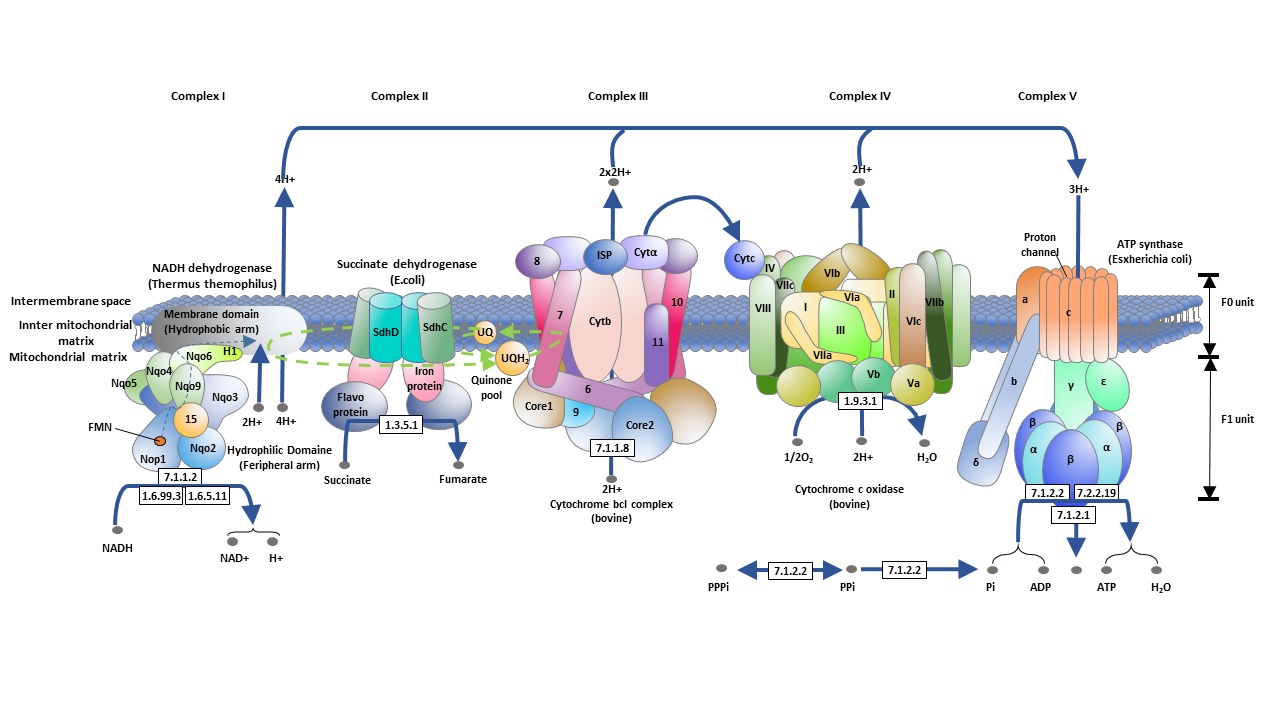
The electron transport chain consists of a series of protein complexes embedded on the inner mitochondrial membrane.
The first functional unit of the electron transport chain is NADH-CoQ oxidoreductase, also named NADH dehydrogenase or Complex I, which serves as the initial site for proton translocation across the membrane. This complex catalyzes NADH oxidation with the assistance of coenzyme Q10. When two electrons pass through Complex I, four hydrogen protons are transported from the mitochondrial matrix into the intermembrane compartment.
Succinic-coenzyme Q oxidoreductase, also known as succinate dehydrogenase or Complex II, is the second component of the ETC. It catalyzes the oxidation of succinate into fumarate and the reduction of coenzyme Q10 to ubiquinol (QH₂). This reaction does not involve transmembrane electron transport or outward proton pumping, and releases far less free energy compared to NADH oxidation. A third proton translocation entry point on the electron transport chain is electron transfer flavin-coenzyme Q oxidoreductase (electron transfer flavin dehydrogenase), which reduces Q10 using electrons from electron transfer flavoproteins located in the mitochondrial matrix.
Coenzyme Q-cytochrome C reductase, or Complex III, catalyzes the oxidation of QH₂ and the reduction of cytochrome c and ferritin. Cytochrome c acts as a mobile single-electron carrier. Coenzyme Q is reduced to QH₂ on one side of the mitochondrial membrane, while QH₂ is oxidized back to CoQ10 on the opposite side; this cyclic electron circulation facilitates transmembrane proton transport and strengthens the proton concentration gradient.
Cytochrome c oxidase (Complex IV) is the last protein complex in the electron transport chain. It completes the terminal reaction of the ETC: transferring electrons to oxygen, the ultimate electron acceptor. Oxygen is reduced to water during this process, accompanied by active proton pumping across the membrane. Both protons directly pumped by this complex and those consumed during oxygen’s reduction to water further amplify the transmembrane proton gradient.
In the final step, the proton-motive force built by the hydrogen ion concentration gradient propels ATP synthase to phosphorylate ADP and generate ATP.
Eukaryotic cells contain another electron-donating molecule called FADH₂, an intermediate metabolite produced in early respiration stages including glycolysis and the tricarboxylic acid cycle. Due to its lower electron transfer potential relative to NADH, FADH₂ skips Complex I and delivers electrons to the ETC through Complex II. During this reaction, FADH₂ is oxidized back to FAD, and coenzyme Q is reduced to QH₂; this reaction also fails to pump protons outward. All subsequent biochemical steps are nearly identical to those in the NADH-initiated electron transport chain.
Prokaryotes like bacteria and archaea possess abundant electron transfer enzymes that can utilize an extremely wide range of chemical substances as substrates. Similar to eukaryotic cells, prokaryotic electron transport uses energy released from substrate oxidation to pump protons across the cytoplasmic membrane, creating an electrochemical gradient that drives ATP synthase to produce ATP. The core difference is that bacteria and archaea can utilize numerous unique compounds as electron donors or acceptors, allowing them to survive and reproduce in diverse environmental habitats.
Under normal physiological conditions, electron transport and ATP phosphorylation remain tightly coupled. Certain chemical compounds disrupt electron transfer or interfere with phosphorylation reactions, leading to abnormal oxidative phosphorylation activity. Four major influencing factors are introduced below:
- Respiratory chain inhibitors: These substances block electron transmission at specific positions on the respiratory chain and suppress oxidation reactions. Compounds including rotenone, phenoxymycin A, barbital and ampicillin bind iron-sulfur proteins within NADH-Q reductase, cutting off electron flow from NADH to CoQ. Antimycin A and dimercaptopropanol hinder electron transfer between Cytb and Cytc1. Cyanide, azide, hydrogen sulfide and carbon monoxide inhibit cytochrome oxidase, preventing electrons from being delivered to oxygen.
- Oxidative phosphorylation inhibitors: These reagents directly interfere with ATP formation and indirectly block electron transport. Oligomycin and dicyclohexylcarbonyldiimide bind to the F0 subunit of ATP synthase, blocking proton backflow through the transmembrane channel. This interrupts the phosphorylation process and fully inhibits oxidative phosphorylation in intact mitochondria.
- Uncoupling agents: These substances separate the two linked processes of electron transport and ATP synthesis. They only suppress ATP production without affecting electron transfer activity. Energy released from electron transport is converted into thermal energy instead of chemical energy, causing excessive consumption of oxygen and metabolic substrates. A typical representative uncoupler is 2,4-dinitrophenol (DNP). As a lipid-soluble molecule, DNP can freely cross mitochondrial membranes: it releases H⁺ inside the matrix and carries protons back to the intermembrane space, eliminating the transmembrane hydrogen gradient. As a result, energy released from oxidation cannot support ATP synthesis, making DNP a typical mobile proton carrier.
In healthy organisms, the overall rate of oxidative phosphorylation is mainly controlled by ADP concentration. When cellular ATP consumption rises, ADP levels increase; once transported into mitochondria, ADP accelerates the rate of oxidative phosphorylation. On the contrary, insufficient ADP slows down the entire pathway. This regulatory mechanism enables ATP production speed to adapt dynamically to the body’s physiological energy demands.
Thyroid hormones can activate Na⁺-K⁺ ATPase on the cell membrane of most tissues, accelerating the decomposition of ATP into ADP and inorganic phosphate (Pi). This raises the volume of ADP transported into mitochondria, lowers the intracellular ATP/ADP ratio, and speeds up oxidative phosphorylation. As ATP synthesis and decomposition rates increase, the body’s oxygen consumption and heat production rise, elevating the basal metabolic rate. This indicator serves as one of the key clinical diagnostic markers for hyperthyroidism patients.
Mitochondrial DNA (mtDNA) exists as a naked circular double-stranded helix without histone protection and complete damage repair systems, making it highly prone to mutations induced by byproducts of oxidative phosphorylation. mtDNA encodes 13 protein subunits essential to oxidative phosphorylation complexes. Therefore, mtDNA mutations disrupt the oxidative phosphorylation workflow, reduce cellular ATP output, and trigger a series of related metabolic disorders.
Oxidative phosphorylation undertakes multiple vital physiological functions within the human body, so any functional defect of this pathway will directly induce disease.
Most mitochondrial disorders are associated with impaired oxidative phosphorylation. Tissues with high energy demand, such as the brain, peripheral nerves, retina, skeletal muscle and cardiac muscle, are particularly susceptible to OXPHOS dysfunction. When oxidative phosphorylation malfunctions in these tissues, clinical symptoms may include epileptic seizures, hypotonia, ophthalmoplegia, convulsions, muscle weakness and cardiomyopathy.