
ELISA stands for Enzyme‑Linked Immunosorbent Assay. It is a qualitative and quantitative detection technique combining solid‑phase immunoassay, specific antigen‑antibody immune reaction and enzyme‑catalyzed color‑generating biochemical reaction. Featuring simple operation, high‑throughput capability, excellent sensitivity and cost‑effectiveness, ELISA serves as a routine core assay in life‑science basic research, clinical diagnosis, animal epidemic detection, food safety screening and biomedical R&D. It is an essential experimental skill for research and laboratory staff. Compared with Western Blot and flow cytometry, ELISA is more suitable for quantitative screening of batch samples with broader application scenarios and better practicality.
The fundamental logic of ELISA relies on reversible specific antigen‑antibody binding, which accounts for its high specificity and low interference. Antigen‑antibody binding is highly specific: antibodies only recognize corresponding antigen epitopes and avoid non‑specific binding with miscellaneous proteins, homologues and impurities in samples, ensuring detection accuracy. The assay system consists of two key modules: the immune recognition module for specific capture and binding of target analytes; and the enzyme‑mediated signal amplification module. Through high‑efficiency catalysis by labeled enzymes, faint molecular binding events are converted into visible and instrument‑readable optical color signals, enabling detection of trace analytes that cannot be directly measured by ordinary instruments.
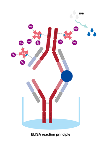
Standard quantitative ELISA follows a rigorous standardized workflow from solid‑phase coating to final readout. Deviations in any step will directly affect experimental results. The general workflow: Coating → Blocking → Sample loading → Antibody incubation → Enzyme conjugation → Color development → Stopping → OD measurement. The whole procedure can be divided into four phases: solid‑phase preparation, immune binding, signal color development and data readout. Below is a detailed breakdown of function, conditions and key notes for each step.
1. Coating: Purified specific antigen or capture antibody is physically adsorbed onto the bottom of polystyrene microplate wells. Typical coating conditions: 4 °C overnight or 37 °C for 2 h. Protein concentration and buffer pH should be well‑controlled for stable adsorption and preserved protein activity. Coating forms the experimental foundation; uneven coating or protein inactivation leads to irregular color development and large numerical deviation.
2. Blocking: After coating, numerous unoccupied hydrophobic sites remain on microplate surfaces. Common blocking reagents include 5 % skim‑milk powder and BSA solution. Standard blocking conditions: 37 °C for 1 h or 4 °C overnight. Blocking saturates vacant sites to minimize non‑specific interference.
3. Sample loading: Discard blocking solution and perform plate washing. Add serially diluted standards, test samples, blank controls and negative controls. Set 2‑3 replicate wells for each group to reduce experimental error. Pipette precisely, avoid bubbles and well‑wall contamination. Follow recommended sample dilution factors to prevent hook effect caused by samples exceeding detection ranges.
4. Antibody incubation: Seal the microplate and incubate at constant 37 °C. Incubation temperature and duration determine binding efficiency. Insufficient temperature or short incubation leads to inadequate antigen‑antibody binding, weak color and low readings. Excessively high temperature or prolonged incubation causes non‑specific binding and protein denaturation. Thoroughly wash wells after incubation to remove unbound sample and impurities.
5. Enzyme conjugation: Add HRP‑ or AP‑labeled specific secondary antibody and incubate to allow binding to immune complexes inside wells. This step anchors enzyme molecules and realizes signal amplification. Complete plate washing is required afterwards to remove unbound free enzyme‑conjugated antibodies.
6. Color development: Add chromogenic substrate under light‑shielded conditions and incubate at 37 °C for 15 min. Enzyme conjugates catalyze redox reactions of substrates to produce soluble colored products. Within valid detection ranges, higher target analyte concentration brings more bound enzyme molecules, generating more colored products and deeper color. Light exposure triggers spontaneous substrate coloration and high background, so light protection is mandatory.
7. Stopping and OD measurement: Add stop solution immediately once incubation finishes to terminate enzyme‑catalyzed reactions and stabilize color signals, avoiding over‑development and value drift. Read OD absorbance values with a microplate reader at corresponding maximum wavelength (450 nm for TMB substrate). Target analyte concentrations of test samples are calculated by fitting against standard curves.
ELISA cannot directly visualize or measure biomolecules. All experimental readings originate from three cascaded reactions: specific immune recognition → enzyme‑mediated signal amplification → optical signal conversion. This cascade grants ELISA superior sensitivity: faint molecular interactions are amplified hundreds‑ to thousands‑fold by enzymatic catalysis for instrument capture.
First comes specific immune binding: immobilized capture antibodies or antigens on microplates capture target analytes from samples and form solid‑phase immune complexes. Unbound impurities and miscellaneous proteins are eliminated in washing steps to guarantee specific signal sources. Next is enzyme labeling and immobilization. Two major reporter enzymes are applied: HRP (Horseradish Peroxidase, most widely‑used in commercial kits) and AP (Alkaline Phosphatase, higher sensitivity for ultra‑trace detection). Enzyme‑conjugated antibodies bind immune complexes and enrich reporter enzymes at target sites.
Then enzyme‑catalyzed color amplification takes place. After substrate addition, anchored enzymes continuously drive chemical reactions and produce stable colored products. Higher target analyte concentration corresponds to more enriched enzymes, higher concentration of colored products and darker solution color, and vice versa. Thanks to this amplification mechanism, ELISA detects analytes down to ng/mL or pg/mL levels, outperforming conventional physicochemical assays.
Finally optical quantization is performed. Microplate readers convert subjective visual color intensity into objective OD absorbance values via photoelectric components. Within valid linear ranges of kits, OD values positively correlate with target concentrations (except competitive ELISA). Absolute concentrations of unknown samples are calculated by fitting standard‑concentration‑OD datasets into standard curves, achieving precise quantitative measurement from qualitative observation. Numerical data can be archived and reviewed to satisfy research traceability and reproducibility requirements.
ELISA depends on intact antigen epitopes for antibody binding, so it mainly detects large biomolecules with complete immune‑binding sites across research, clinical and testing fields. Detectable analytes include functional proteins, structural proteins, specific antigens, pathogen‑specific antibodies, cytokines (IL‑6, TNF‑α, etc.), inflammatory markers, tumor markers, hormones, growth factors, therapeutic proteins and food‑safety toxin proteins. In principle, any macromolecule with well‑matched paired antibodies can be quantified with ELISA.
ELISA accepts diverse sample matrices: clinical specimens (human serum, plasma, urine, cerebrospinal fluid, pleural effusion), research samples (cell culture supernatant, animal serum, tissue homogenate, cell lysate), food and environmental samples (food extracts, water extracts, feed samples). Important limitations: small‑molecule substances (small‑molecule hormones, veterinary drug residues, mycotoxins) lack multiple epitopes for sandwich assay and must be tested with competitive ELISA. Unknown substances without specific antibodies and small inorganic molecules cannot be measured by ELISA, a common pitfall for beginners.
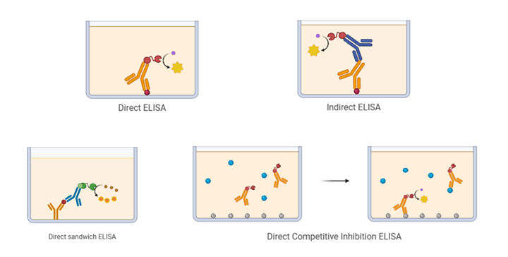
Based on coating strategies, immune‑binding modes and molecular weight of targets, ELISA falls into four classic formats. They differ drastically in experimental logic, applicable analytes, value trends and pros‑cons. Researchers must select proper formats according to analyte molecular weight and experimental purpose; wrong selection will lead to complete assay failure.
1. Direct ELISA
Principle: Enzyme‑conjugated primary antibody directly binds coated antigens on microplates; immune binding and enzyme labeling are completed in one step without secondary antibody. Advantages: simple workflow and short assay duration with minimal step‑induced errors. Drawbacks: limited signal amplification leads to low sensitivity and specificity with frequent non‑specific binding. Custom enzyme‑labeled primary antibodies for every target result in poor versatility. Application: only for rapid qualitative screening of simple antigens rather than precise quantification. Rarely adopted in formal research and clinical tests, mostly used for teaching demonstration.
2. Indirect ELISA
Principle: Purified antigens are coated on wells to capture primary antibodies from samples. Universal enzyme‑labeled secondary antibodies then bind the Fc fragment of primary antibodies. Advantages: excellent versatility and high sensitivity. One type of labeled secondary antibody works for multiple primary antibodies to reduce cost; dual‑layer immune binding improves specificity. Application: exclusively for antibody detection, such as serum antibody screening for viral infection, vaccine titer measurement and autoantibody detection. Preferred format for antibody assays and not suitable for antigen measurement.
3. Sandwich ELISA
Principle: Immobilized capture antibodies grab target antigens, then detection antibodies bind another epitope of antigens to build a "capture antibody‑antigen‑detection antibody" sandwich complex, followed by enzyme‑conjugated secondary antibody for color development. It is the most widely‑used ELISA format with high accuracy and good reproducibility. Advantages: dual‑antigen‑antibody recognition minimizes non‑specific interference, delivers top‑tier sensitivity and low quantitative error. Application: detection of macromolecular antigens (> 5 kDa) including cytokines, protein biomarkers and antibodies. Suitable for most research quantitative assays and clinical biomarker tests. Value trend: OD values rise with increasing antigen concentration (positive correlation).
4. Competitive ELISA
Principle: Designed for small molecules lacking multiple epitopes for sandwich configuration. Sample small‑molecule analytes compete with enzyme‑labeled reference antigens for limited binding sites of immobilized antibodies. Features and value trend are opposite to sandwich ELISA: higher target concentration in samples occupies more antibody sites, less labeled antigen binds to wells, color gets weaker and OD value decreases. Application: small‑molecule detection including hormones, veterinary drug residues, mycotoxins, pharmaceutical small molecules and metabolites. Mainstream format for food‑safety and drug‑residue screening.
Core strengths (Why ELISA remains a mainstream detection technique)
1. High specificity: Antibodies recognize target‑specific epitopes and resist interference from miscellaneous proteins and homologues. Low false‑positive and false‑negative rates deliver reliable results that satisfy rigorous standards for research and clinical diagnosis.
2. Outstanding sensitivity: Enzymatic signal amplification enables measurement of trace analytes at pg/mL‑ng/mL ranges, capturing subtle concentration changes of low‑abundance targets such as cytokines and minor hormones.
3. High throughput and efficiency: Standard 96‑well / 384‑well microplates support dozens‑to‑hundreds samples in single runs. Combined with automated plate washers and microplate readers, ELISA suits large‑scale animal studies, clinical sample screening and bulk food inspection.
4. Low‑cost and accessible: Only basic incubator and microplate reader are required without high‑end instruments or complicated maintenance. Commercial ELISA kits are cost‑effective and pre‑formulated to lower technical barriers.
5. Broad compatibility: Accepts specimen matrices from body fluids, cells, tissues to food samples. Applications cover basic research, clinical medicine, veterinary science, food safety, biomedicine and environmental testing. Quantitative, repeatable and traceable output meets standards for research papers and test reports.
Main limitations (Key pitfalls for experimental avoidance)
1. Heavily dependent on antibody quality: Antibody specificity, purity and activity determine assay success. Poor‑specificity antibodies with cross‑reactivity cause high background and false positives. Degraded antibody activity produces weak color signals, low readings or blank results, which is the leading cause of ELISA failure.
2. Target‑dependent detection only: ELISA is a targeted assay. It can only detect known analytes with validated antigen‑antibody pairs. Unknown analytes without corresponding antibodies cannot be screened, so it cannot perform omics‑style untargeted profiling.
3. Hook effect interference: Excessively high antigen concentration causes saturation and disturbs immune equilibrium, leading to weakened color signals and underestimated measured concentrations. Beginners frequently encounter distorted data and invalid standard curves for undiluted high‑concentration samples.
4. Vulnerable to manual operation errors: Incubation temperature and duration, washing cycles and intensity, pipetting accuracy, light shielding and stopping timing all impact results. Improper operations bring high well‑to‑well CV values, plate‑to‑plate variations and poor reproducibility, demanding skilled operators.
5. Limited accuracy for small‑molecule assays: Sandwich ELISA delivers highly precise quantification for macromolecules. Competitive ELISA for small molecules shows inferior stability and specificity, higher background noise and larger relative errors. It cannot distinguish structurally‑similar small‑molecule homologues.